Have you ever wondered how a simple movement, like lifting a glass of water, is possible? It involves a fascinating interplay of complex structures within our bodies, particularly the intricate world of skeletal muscle. This muscle type, responsible for our voluntary movements, is a masterpiece of nature, built with a precise architecture that allows for efficient force generation. In this article, we’ll take a deep dive into the microscopic world of skeletal muscle, exploring its anatomy and organization. Understanding these structures will shed light on the mechanisms behind muscle function and how we move.

Image: phoebe-chapter.blogspot.com
Skeletal muscle, like other tissues in the body, is hierarchically organized, with each level building upon the previous one. This structure is essential for enabling the muscle to contract efficiently and generate the force needed to perform our daily activities. We will journey from the largest structural units down to the molecular level, unveiling how each component plays a vital role in muscle activity.
A Glimpse into the Muscular Architecture
The Building Blocks: Muscle Fibers
At the most macroscopic level, skeletal muscle appears as a collection of bundles, known as fascicles, arranged in a parallel fashion. Each fascicle is further subdivided into individual muscle fibers, the fundamental units of muscle tissue. A single muscle fiber, in essence, is a multinucleated cell, meaning it houses multiple nuclei within its cytoplasm. These nuclei are responsible for controlling protein synthesis within the fiber, playing a crucial role in muscle growth and repair.
The Microscopic World of Sarcomeres
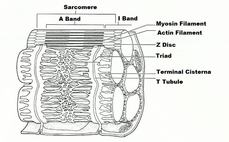
Within each muscle fiber, we discover the fascinating world of sarcomeres, the basic functional unit of muscle contraction. Sarcomeres are arranged in a repeating pattern along the length of the fiber, resembling tiny compartments. Each sarcomere consists of highly organized protein filaments: actin and myosin. Actin, the thinner filament, forms a double helix, while myosin, the thicker filament, is composed of numerous elongated proteins with globular heads.

Image: mavink.com
The Sliding Filament Theory: A Symphony of Movement
The magic of muscle contraction lies within the interplay between these protein filaments. The sliding filament theory explains how muscle contraction occurs. Briefly, the myosin heads attach to the actin filaments and pull them towards the center of the sarcomere, causing the sarcomere to shorten. This shortening of numerous sarcomeres collectively produces the contraction of the entire muscle fiber.
The Role of the Sarcoplasmic Reticulum (SR)
The smooth endoplasmic reticulum of muscle cells, known as the sarcoplasmic reticulum (SR), plays a vital role in calcium regulation. Calcium ions are crucial for initiating muscle contraction by binding to troponin, a protein associated with the actin filament. This binding triggers a conformational change in troponin, exposing active sites on the actin filament to which the myosin heads can bind, initiating the sliding filament process.
Nerve Control: The Role of Motor Neurons
Skeletal muscle is under voluntary control, meaning we can consciously activate it. This control is achieved through the nervous system. Motor neurons, specialized cells in the nervous system, transmit signals from the brain and spinal cord to muscle fibers. When a motor neuron sends a signal to a muscle fiber, it triggers the release of acetylcholine, a neurotransmitter that binds to receptors on the muscle fiber’s surface. This binding event initiates a series of events leading to the release of calcium from the SR, ultimately resulting in muscle contraction.
Beyond Muscle Fibers: Organizational Structures
The remarkable organization of skeletal muscle doesn’t end at the level of muscle fibers. These fibers are arranged into larger structures, ensuring efficient force transmission and coordinated contraction.
Fascicles: Bundles of Strength
We’ve already mentioned that muscle fibers are grouped into bundles called fascicles. Each fascicle is enveloped by a connective tissue sheathing called perimysium. This sheathing serves to hold the fibers together, allowing for coordinated contraction within the fascicle and providing pathways for blood vessels and nerves to reach individual fibers.
The Whole Muscleg: From Fiber to Function
Numerous fascicles are then bundled together to form a complete muscle, covered by a dense connective tissue layer known as epimysium. This outer layer is essential for maintaining the muscle’s shape and for attaching it to bones via tendons. The epimysium also helps to transmit the force generated by individual muscle fibers to the tendons, enabling movement of the entire limb or body part.
The Importance of Microscopic Anatomy in Exercise
Understanding the microscopic anatomy of skeletal muscle is not merely an academic pursuit. It has significant implications for exercise and fitness. The organization of muscle fibers, sarcomeres, and the intricate mechanisms of contraction directly influence how muscles respond to training.
Muscle Adaptations: The Strength of Training
When we engage in exercise, particularly strength training, our muscle fibers undergo adaptation. These adaptations involve an increase in the size and number of muscle fibers, leading to enhanced muscle strength and power. This process, known as hypertrophy, is driven by the body’s response to the increased demands placed upon the muscles.
The Role of Microscopic Adaptations in Performance
Understanding the microscopic adaptations that occur in response to exercise can guide our training strategies. Targeting specific muscle fiber types can enhance performance in different types of activities. For example, endurance training primarily affects slow-twitch muscle fibers, while weight training primarily targets fast-twitch muscle fibers.
Beyond Muscle Growth: Improved Health
Beyond enhancing strength and performance, understanding the microscopic anatomy of skeletal muscle is vital for promoting overall health. Muscle tissue plays a critical role in maintaining blood sugar levels, burning calories, and even bolstering the immune system. Exercise, by stimulating muscle growth and function, contributes to a healthier and more resilient body.
Exercise 11: A Deep Dive into Skeletal Muscle
“Exercise 11” often refers to a laboratory exercise in an anatomy or physiology course. Such exercises typically involve examining skeletal muscle under a microscope to visualize the structures discussed above. Students might use prepared slides or even sections of fresh muscle tissue to observe the arrangement of muscle fibers, fascicles, and sarcomeres. This hands-on experience helps solidify their understanding of the microscopic anatomy of skeletal muscle and its significance in overall body function.
Exercise 11 Microscopic Anatomy And Organization Of Skeletal Muscle
Conclusion: Unveiling the Power of Microstructure
The intricacies of skeletal muscle organization and function, from the smallest sarcomere to the largest muscle, illustrate the remarkable complexity and efficiency of our bodies. By understanding the microscopic anatomy of skeletal muscle, we gain a deeper appreciation for how our movements are generated and how exercise can shape and refine these remarkable structures. Whether you’re an athlete looking to optimize performance or simply interested in understanding the mechanics of your own body, delving into the microscopic world of skeletal muscle provides valuable insights into the foundation of movement and the power of our own biological design.


![Cyclomancy – The Secret of Psychic Power Control [PDF] Cyclomancy – The Secret of Psychic Power Control [PDF]](https://i3.wp.com/i.ebayimg.com/images/g/2OEAAOSwxehiulu5/s-l1600.jpg?w=740&resize=740,414&ssl=1)

